An Illustrated Guide to Oral Histology
€128,00 €116,00
Learn more about the histological presentation of diseased and normal oral tissues with this high definition illustrated dental reference!
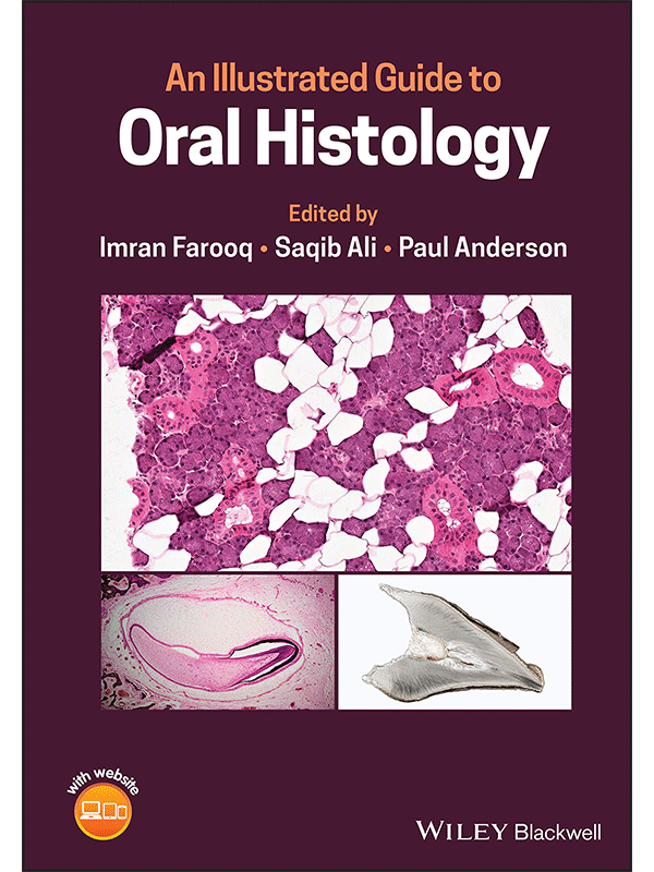
An Illustrated Guide to Oral Histology delivers a collection of high-definition histological and pathological images, presenting both diseased and normal oral tissues.
The book provides over 200 high-magnification histomicrographs of oral tissues, as well as definitions and explanations of key identifying histological and pathological features of oral tissues. Readers will also benefit from explanations of the clinical significance of particular features, numerous images of ground sections, haemotoxylin- and eosin-stained sections, and electron images. It also includes core topics such as:
- An introduction to tooth development, including the bud, cap, early bell, and late bell stages
- A thorough exploration of enamel, dentin, cementum and dental pulp
- A discussion of the periodontal ligament, including alveolar crest fibers, horizontal, oblique, apical, and inter-radicular fibers, transseptal fibers, and gingival fibers
- A guide to alveolar bone, oral mucosa, and salivary glands
Perfect for postgraduate dental students, An Illustrated Guide to Oral Histology will also be useful to undergraduate dental students, and those looking to improve their understanding of the microscopic structure of dental tissues and their pathologies.
Δείτε τα Περιεχόμενα
Δείτε Απόσπασμα του Βιβλίου
Διαθέσιμο κατόπιν παραγγελίας





Αξιολογήσεις
Δεν υπάρχει καμία αξιολόγηση ακόμη.